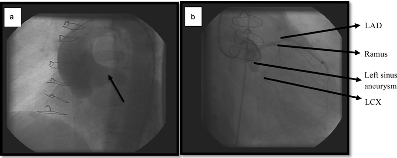
Fig. 1.

(a) Aortogram showing the aneurysm of the left sinus of Valsalva. (b) Attempted coronary angiography showing the left sinus aneurysm with coronary arteries at the end LAD and LCX arteries at the end. LAD, left anterior descending; LCX, left circumflex.
