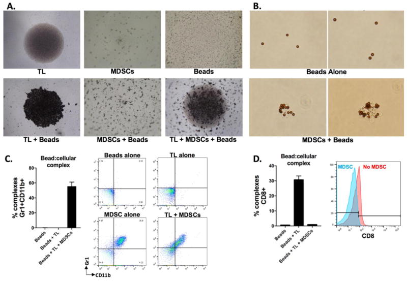
Figure 2. MDSCs sequester CD3/28 microbeads and prevent their contact with T-lymphocytes.
A, 4x photomicrographs of round-bottom wells containing the indicated cell type(s) and/or CD3/28 microbeads after 24 hours in culture. All components were added to culture in equal ratios. B, 40x photomicrographs of CD3/28 microbeads cultured for 24 hours alone or in the presence of MDSCs at a 1:1 ratio. C&D, CD3/28 microbeads and T-lymphocytes with or without MDSCs as indicated were co-cultured in equal ratios for 3 days. Microbeads were then magnetically isolated, and microbead:cellular complexes were stained with fluorophore conjugates antibodies as indicated and analyzed via flow cytometry. Left panels, quantification of complexes positive for markers as indicated, right panels, representative dot plots or histograms. All results shown are representative of a least 3 independent assays. TL, T-lymphocytes.

