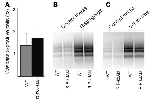
Figure 7.
Assessment of islet apoptosis in RIP-kdAkt and WT mice. (A) Frequency of activated caspase-positive cells in islets from RIP-kdAkt and WT mice (n = 3). (B) DNA laddering in RIP-kdAkt and WT islets after 48 hours of incubation in RPMI containing 10 mM glucose, 10% serum (control medium), and 1 μM thapsigargin. (C) Apoptosis assessment by DNA laddering in RIP-kdAkt and WT islets cultured for 7 days in control medium and in serum-free RPMI with 10 mM glucose. Data are representative of at least 3 experiments in duplicate.

