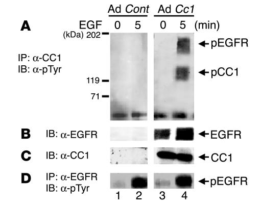
Figure 2.
EGFR phosphorylates CEACAM1-4L in infected human MDA-MB468 breast cancer cells. MDA-MB468 breast cancer cells were infected with Ad Cc1 (lanes 3 and 4) or with Ad Cont (lanes 1 and 2) prior to EGF treatment. Equal amounts of proteins were immunoprecipitated with α-CC1 (A) or α-EGFR (D) prior to Western blot analysis with α-pTyr. (B) Proteins in A were reprobed with α-EGFR. (C) To account for the amount of CEACAM1-4L, amounts of proteins equal to those in A were applied on the same SDS-PAGE gel and were immunoblotted with α-CC1. The results obtained were consistent in at least 3 experiments.

