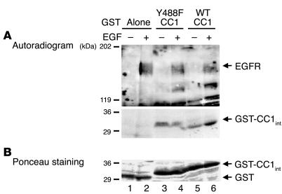
Figure 3.
CEACAM1-4L is a direct substrate of EGFR. (A) After treatment of MDA-MB468 cells with EGF (100 nM) (+) or buffer alone (_) for 5 minutes, cells were lysed and EGFR was extracted by immunoprecipitation with α-EGFR. GST fusion to intracellular peptides of wild type (WT CC1; lanes 5 and 6) or Y488F mutant (Y488F CC1; lanes 3 and 4) CEACAM1-4L (GST-CC1int) were incubated with equal amounts of EGFR immunoprecipitates in the presence of [γ-32P]ATP for 10 minutes prior to the addition of SDS buffer and analysis by 6_12% SDS-PAGE and autoradiography for the detection of phosphorylated proteins. Peptide-free GST was included as control to account for nonspecific association (Alone; lanes 1 and 2). (B) The amount of GST peptides from the experiment in A, measured by Ponceau S staining. The results obtained were consistent in at least three experiments. The two gels in A are from the same SDS-PAGE gel and differ in the exposure time of the autoradiogram for optimal visualization of both EGFR and GST bands.

