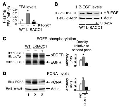
Figure 8.
Elevated adipokines activate EGFR activation in L-SACC1 hepatocytes. Five age-matched male mice were treated with KT6-207 (+; black bars) or vehicle alone (_; gray bars for L-SACC1 mice and white bars for WT mice). (A) At the end of the treatment, blood was removed for determination of plasma FFA levels. (B) Visceral adipose tissues were removed from the intra-abdominal cavity and lysed for sequential immunoblotting with α_HB-EGF followed by α-actin for determination of HB-EGF content. (C) Livers were removed from age-matched mice and lysed and equal amounts of proteins were subjected to immunoprecipitation with α-EGFR prior to sequential immunoblotting with α-pTyr for examination of EGFR phosphorylation and α-EGFR to account for the amount of this protein in the immunoprecipitates. (D) Equal amounts of proteins were analyzed by Western blotting with α-PCNA and were normalized by reprobing with α-actin for determination of cell proliferation. *P < 0.05 versus (_) WT.

