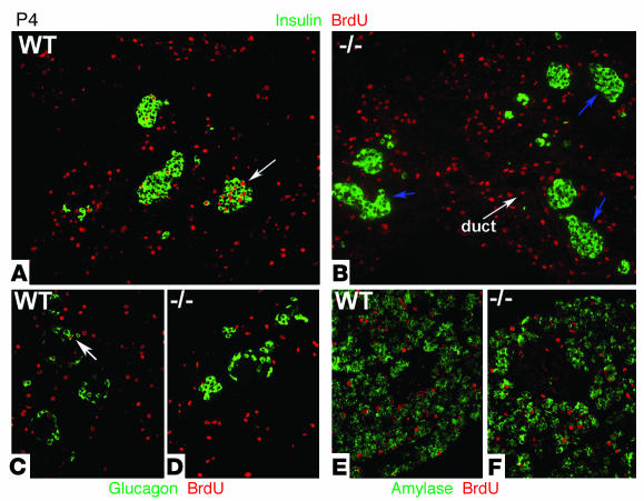
Figure 3.
Incorporation of BrdU during postnatal pancreatic development of WT and cyclin D2–/– mice. BrdU was injected in 4- and 7-day-old mice 2 hours before being sacrificed. (A and B) Sections from pancreata costained with anti_insulin and anti_BrdU Ab’s. (A) In P4 WT mice, a fraction of β cells that incorporate BrdU are evident during the first week of postnatal development. Quantification of 20 representative islets showed that 9.2% of β cells incorporated BrdU in P4 WT mice. Arrow indicates an example of an islet with BrdU-positive β cells. (B) In cyclin D2–/– littermates, β cells that incorporated BrdU are not observed. Arrows indicate islets in cyclin D2–/– that do not contain BrdU-positive β cells. (C and D) Sections from pancreas costained with anti_glucagon and anti_BrdU Ab’s. (C) Glucagon-positive cells incorporate BrdU in the WT pancreas. Arrow indicates an example of an islet with a BrdU-positive α cell. (D) No glucagon-positive cells that incorporate BrdU are observed. (E and F) Sections from pancreas costained with anti_amylase and anti_BrdU Ab’s. BrdU incorporation is similar in exocrine and ductal tissue of the pancreata from WT (E) and cyclin D2–/– mice (F).

