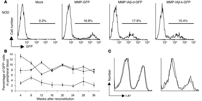
Figure 3.
Expression of retrovirally transduced MHC class II β chains in vivo. (A) MMP-IAβ-d-GFP and MMP-IAβ-k-GFP viruses effectively infect primary bone marrow cells. Bone marrow cells were harvested from 5-fluorouracil_treated NOD mice and transduced as described. Immediately after transduction, bone marrow cells were harvested and examined for GFP expression by flow cytometry. One representative experiment of 5 independent experiments is shown. (B) Expression of retrovirally transduced I-Aβ chains is stable. Expression of GFP in PBMCs of NOD mice reconstituted with MMP-IAβ-d-GFP_transduced (squares), MMP-IAβ-k-GFP_transduced (triangles), or control MMP-GFP_transduced bone marrow (diamonds), determined by flow cytometry. Shown are the mean values obtained for 5_6 mice per time point. The data are representative of 3 independent experiments. (C) MHC class II expression levels on the surface of PBMCs. Twenty weeks after bone marrow transplantation, PBMCs were harvested from mice reconstituted with bone marrow transduced with either MMP-IAβ-k-GFP (solid line) or control MMP-GFP virus (dashed line). Cells were stained with anti_I-Ad antibody 39-10-8, which recognizes all cell surface MHC class II, and analyzed by flow cytometry. Left panel: Total cells. Right panel: GFP+ transduced cells.

