Figure 4.
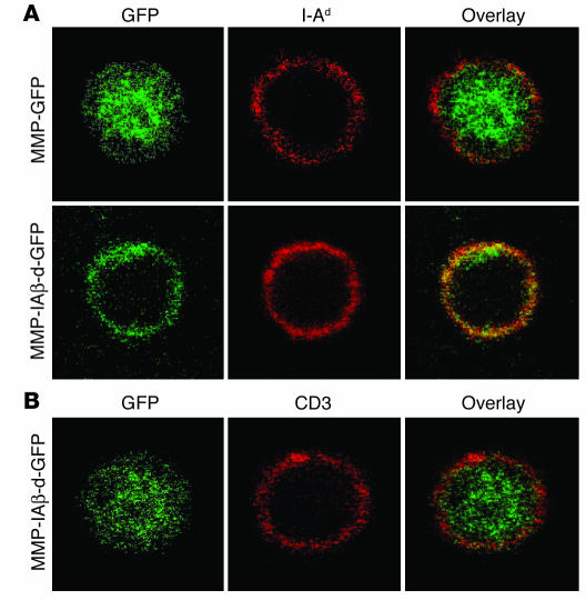
Subcellular localization of retrovirally encoded I-Aβ chains. (A) Retrovirally encoded I-Aβ chains are expressed on the surface of MHC class II_positive cells. PBMCs were harvested from NOD mice reconstituted with either MMP-GFP_transduced (top panels) or MMP-IAβ-d-GFP_transduced bone marrow (bottom panels), fixed, and stained with antibodies specific for GFP (left panels, green) and I-Ad (middle panels, red). The anti_I-Ad antibody used cross-reacts with I-Ag7. Right panels: Overlay images showing colocalization of retrovirally encoded I-Ad_GFP fusion proteins with endogenous I-Ag7 on the surface in yellow. (B) Retrovirally encoded I-Aβ chains are expressed in the cytoplasm of surface-MHC class II negative cells. PBMCs from NOD mice reconstituted with MMP-IAβ-d-GFP_transduced bone marrow were fixed and stained with antibodies specific for GFP (left panel, green) and anti-CD3 (middle panel, red). An overlay of the 2 images is shown in the right panel, demonstrating that I-Aβd_GFP does not colocalize with CD3 on the cell surface of MHC class II_negative cells. Shown are representative results of 3 independent experiments.

