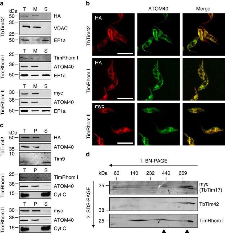
Figure 3. Novel TIM subunits are integral membrane proteins that form a large complex.
(a) Immunoblot analysis of whole-cell (T), digitonin-extracted mitochondria-enriched pellet (M) and soluble (S) fractions of cells expressing c-terminally HA-tagged TbTim42 and cells expressing c-terminally myc-tagged TimRhom II. The top and bottom panels were probed with anti-tag antibodies. The middle panel shows a redecoration of the bottom panel with a custom-made antiserum against TimRhom I, thus the same controls are depicted as in the bottom panel. ATOM40/VDAC and Elongation factor 1-alpha (EF1a) serve as markers for mitochondria and cytosol, respectively. (b) IF analysis of cells expressing the indicated tagged proteins. ATOM40 serves as a mitochondrial marker. Scale bar, 10 μm. (c) Immunoblots of the total (T), pellet (P) and supernatant (S) fractions of a carbonate extraction performed at pH 11.5. Mitochondria-enriched pellets of cells expressing c-terminally HA-tagged TbTim42 and cells expressing c-terminally myc-tagged TimRhom II were used as starting material. The top and bottom panels were probed with anti-tag antibodies. The middle panel shows a redecoration of the bottom panel with a custom-made antiserum against TimRhom I, thus the same controls are depicted as in the bottom panel. ATOM40/VDAC and cytochrome c (Cyt c)/Tim9 serve as markers for integral and peripheral membrane proteins, respectively. (d) Digitonin-solubilized mitochondrial protein complexes were separated in the first dimension by 6–16.5% BN–PAGE and then subjected to denaturing 14% SDS–PAGE in the second dimension. The resulting immunoblots were probed for c-terminally myc-tagged TbTim17 using an anti-myc antiserum as well as for TbTim42 and TimRhom I using custom-made antisera. Arrow heads indicate high molecular weight complexes containing TIM subunits. VDAC, voltage-dependent anion channel.

