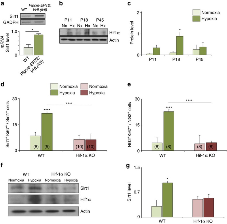
Figure 2. HX-induced Sirt1 expression in white matter OPCs requires HIF1α.
(a) HIF1α stabilization of Sirt1 transcript expression in OPCs as revealed by representative RT–PCR represents higher level of Sirt1 mRNA in VHL cKO mice. GDPDH mRNA serves as a control. Mean±s.e.m., n=3 brains for each group. (b,c) Representative western blot demonstrates a transient increase of HIF1α expression in HX white matter at P18 with no significant effect at P11 (P=0.7955) and P45 (P=0.7333). Histograms show mean±s.e.m. (d,e) Graphs represent the percentages of Sirt1+Ki67+ and NG2+Ki67+ cells after HX white matter in WT and HIF1α KO mice. Number in parentheses within bar indicates number of samples (n=4 brains per group and per genotype; ****P<0.0001, one-way analysis of variance, Bonferroni post hoc test, mean±s.e.m.). (f,g) Western blot demonstrates no increase (P=0.8231) in Sirt1 and HIF1α expression in white matter of Hif1α KO mice. Actin was used as loading control (mean±s.e.m.; n=3 brains for each experiment, group and genotype).

