Figure 11.
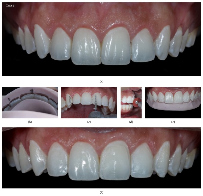
Case 1. (a) Initial view of the teeth before preparation. During treatment, the patient chose to extend the treatment to the bilateral second premolars. (b) Vestibular guide to assess the distance between the dental tissue and the correct placement indicated by the wax model, with information necessary to direct the professional in the decision of whether to wear down the sound tooth structure. (c) Palatal guide to aid the professional in determining the proper cervicoincisal size and the correct positioning of the incisal edge. (d)-(e) Minimally invasive preparations using a sanding disc and diamond burs. (f) Appearance immediately after minimally invasive preparation.

