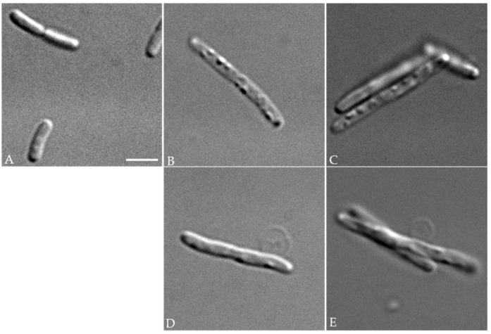
Figure 3.
Morphology of E. coli strains. Optical micrographs of exponentially-growing control FB8 cells (A) and of cells expressing either the wild-type (B,D) or the D222A mutant (C,E) PcaM1. As compared to control cells, those expressing either form of PcaM1 in the periplasm appeared elongated, presented swellings on their surface (B,C), and some of them were blebbing (D,E). The scale bar shown is 3 µm.

