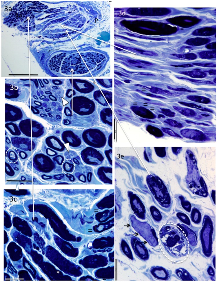
Figure 3.
Methylene- (a–c) and toluidine (d–e) blue-stained sections from AIN. In addition to thickened, split and folded myelin, AIN and PIN nerves showed both transverse (b,e) and longitudinal orientation of axons (c,d). Abnormally pale-stained myelin (possibly uncompacted) can be seen surrounding the typically dark staining myelin (c,d). Note the large demyelinated axons, clearly lacking the myelin component (e) and degenerating vacuolated axons (b) or myelin without an axon (e). Small myelinated (*) and unmyelinated axons (arrowheads) were also observed. Demyelinated axons (small arrows), abnormally pale stained myelin abutting normal myelin (=), degenerating axons (enclosed in white circles). Perineurium (P), long arrows show region of high oower micrographs bar = 200 μm (a), 20 μm (b–e).

