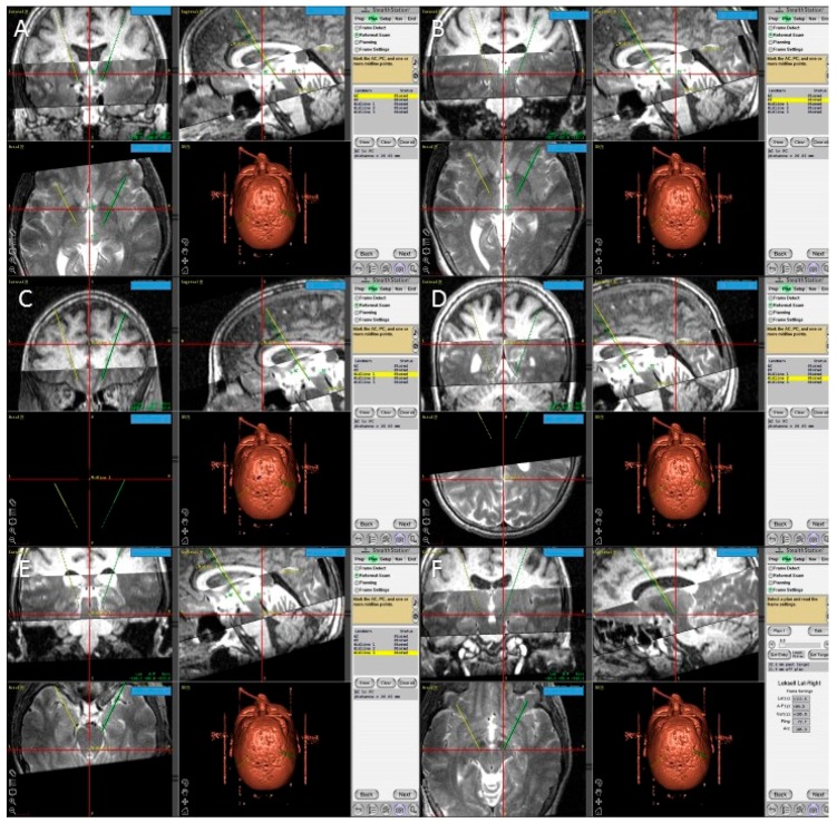
Figure 6.
Screen shots from the FrameLink software of the StealthStation showing fused T1 and T2 magnetic resonance imaging (MRI) images of the patient and the planning process with identification of the posterior edge of the anterior commissure (A); the anterior edge of the posterior commissure PC (B); three midline points (C–E); and the final coordinates of the right subthalamic nucleus (F).

