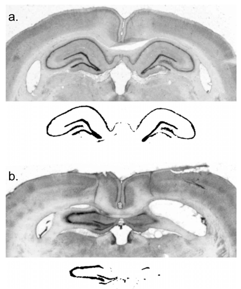
Figure 1.

An index of lesion in each subject was obtained when digital images of the dorsal hippocampus were subjected to a grayscale threshold so that the area within the cell body layers of the surviving dorsal hippocampus was calculated. (a) Grayscale and thresholded images of a control subject; (b) images of a lesioned subject.
