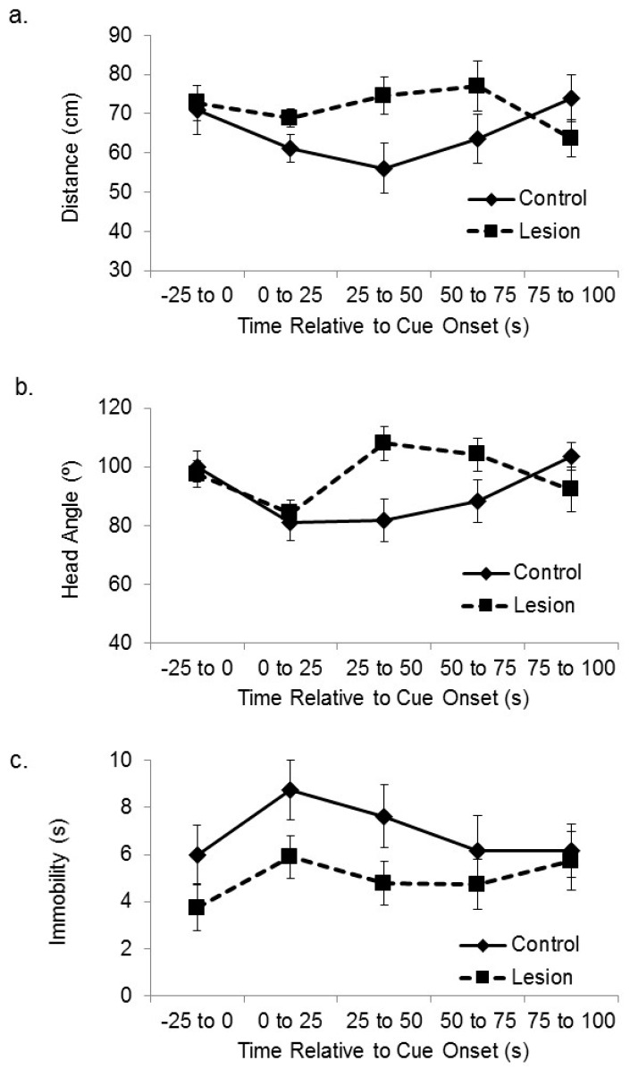
Figure 4.
Medial prefrontal cortex (mPFC) and cue-directed orientation. (a) The distance between the subjects and the corner of the platform nearest the active speaker is plotted during the sensory preconditioning session exposure to the auditory cue and the immediately preceding baseline period. Control subjects approached the active speaker during the cue more than subjects with lesions to the mPFC; (b) The head angle relative to the location of the active speaker during the baseline period and cue exposure is plotted. Control subjects maintained head orientation towards the location of the auditory cue more than subjects with lesions to the mPFC; (c) The number of seconds spent immobile during the baseline period and after cue onset is plotted. The difference between the control and lesioned groups in seconds spent immobile was not statistically significant.

