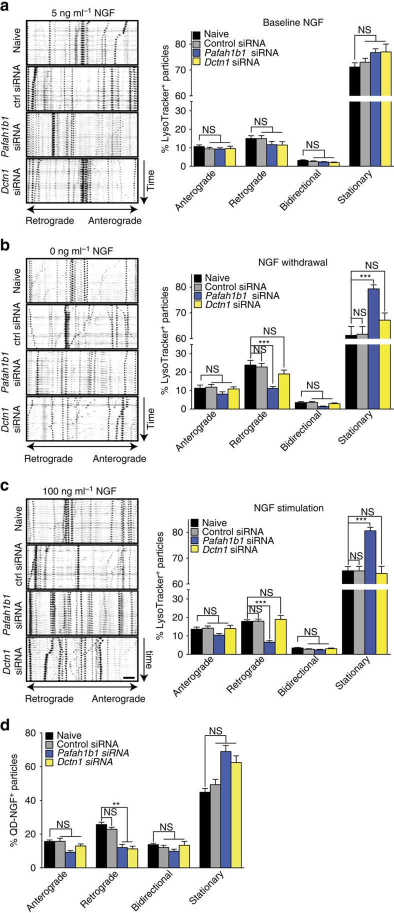
Figure 4. NGF-induced changes in axonal trafficking require local synthesis of Lis1 or p150Glued.
DRG neurons were cultured in microfluidic chambers. On DIV 3, the NGF concentration in the axonal chamber was changed to 5 ng ml−1, and axons were selectively transfected with a non-targeting control siRNA or siRNAs targeting Pafah1b1 or Dctn1. (a–c) After 24 h, fresh medium was added to the axonal chamber containing 5 ng ml−1 NGF, no NGF or 100 ng ml−1 NGF together with LysoTracker Green for 15 min. Live-imaging time-lapse series of axonal fields were acquired, with images being taken every 13 s for 4 min. Kymographs of representative 100-μm-long axonal segments are shown. Scale bar, 10 μm. LysoTracker-positive particles with diameters ≥1 μm were scored as anterograde, retrograde, bidirectional or stationary. Means±s.e.m. of 12–18 optical fields per conditions (n=3–6 biological replicates). **P≥0.01; ***P≥0.001. One-way ANOVA with Bonferroni's multiple comparisons test. (d) On DIV 4, axons were treated with 100 ng ml−1 QD-NGF for 15 min and live imaged as above. QD-labelled particles <1-μm diameter were scored as anterograde, retrograde, bidirectional or stationary. Means±s.e.m. of nine optical fields per conditions (n=3 biological replicates). **P≥0.01; ***P≥0.001. Kruskal–Wallis test with Dunn's multiple comparison test. NS, not significant.

