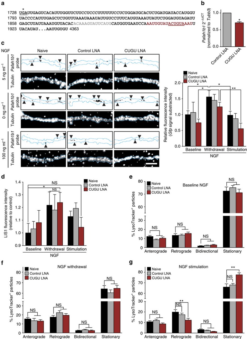
Figure 7. Association with APC separates axonally localized Lis1 transcripts into two functionally distinct pools.
(a) Partial sequence of the 3′-UTR of rat Pafah1b1 starting at the stop codon (*). The binding regions of the CUGU and control LNAs are indicated in maroon and grey, respectively. The CUGU element is underlined. (b) Dissociated DRG were transfected with control and CUGU LNA, and 24 h later, APC RNA immunoprecipitation was performed. Pafah1b1 was quantified by RT–PCR. 2−ΔΔCT values are reported relative to Tubb3 (positive control, binds APC but is not targeted by the LNAs). Gfp was included as a control (no reads detected). Means±s.e.m. (n=2 biological replicates with two technical replicates each). *P≥0.05. t-test. (c) DRG neurons were cultured in microfluidic chambers. On DIV 3, the NGF concentration in the axonal chamber was changed to 5 ng ml−1, and cell bodies were selectively transfected with the control or CUGU LNAs. Twenty-four hours after transfection, axons were treated with 0, 5 or 100 ng ml−1 NGF for 12 h, and axonal Pafah1b1 mRNA levels were determined by FISH. Background fluorescence was determined using a Gfp probe and subtracted. Means±s.e.m. of 15 optical fields per condition (n=3 biological replicates). *P≥0.05. Two-way ANOVA with Fisher's least significant difference test. Scale bar, 5 μm. (d) DRG neurons were cultured and transfected as in a. Twenty-four hours after transfection, axons were treated with 0, 5 or 100 ng ml−1 NGF for 10 min, and axonal Lis1 protein levels were measured by quantitative immunofluorescence. Means±s.e.m. of 20–30 optical fields per conditions (n=4–6 biological replicates). *P≥0.05. Two-way ANOVA with Fisher's LSD test. (e–g) DRG neurons were cultured and transfected as in a. Twenty-four hours after transfection, transport of LysoTracker-positive particles was observed in axons at baseline NGF (e), without NGF (f) or stimulated with NGF (g). Live-imaging time-lapse series of axonal fields were acquired, with images being taken every 13 s for 4 min. LysoTracker-positive particles with diameters ≥1 μm were scored as anterograde, retrograde, bidirectional or stationary. Means±s.e.m. of nine optical fields per conditions (n=3 biological replicates). **P≥0.01. One-way ANOVA with Bonferroni's multiple comparisons test. NS, not significant.

