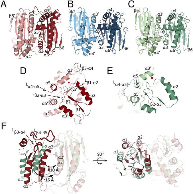
Fig. 3.
Comparison of dimerization among CrLCIB-ΔC, CrLCIC-ΔC and PtLCIB4. (A–C) The two subunits of the PtLCIB4 dimer (red and pale red), CrLCIC-ΔC dimer (blue and pale blue), and CrLCIB-ΔC (green and pale green) are depicted with one zinc ion per subunit. (D and E) The secondary structure elements of PtLCIB4 (D) and CrLCIB-ΔC (E) involved in dimerization are shown. (F) Comparison of CrLCIB-ΔC and PtLCIB4 dimers by alignment based on one subunit (shown in transparency). Secondary structure elements of CrLCIB-ΔC and PtLCIB4 are labeled in dark green and black, respectively. The intersubunit gap distance is measured as the distance between α2 and α2′ and is highlighted by a curved arrow with the distance indicated. The two loops (Lβ3-α4 and Lβ4-β5) that are clearly defined in PtLCIB4 but disordered in CrLCIB/C-ΔC are shown as ribbons with a thicker radius. An alternative view is also depicted with the dimer rotated 90 degrees around an axis perpendicular to the dimeric axis.

