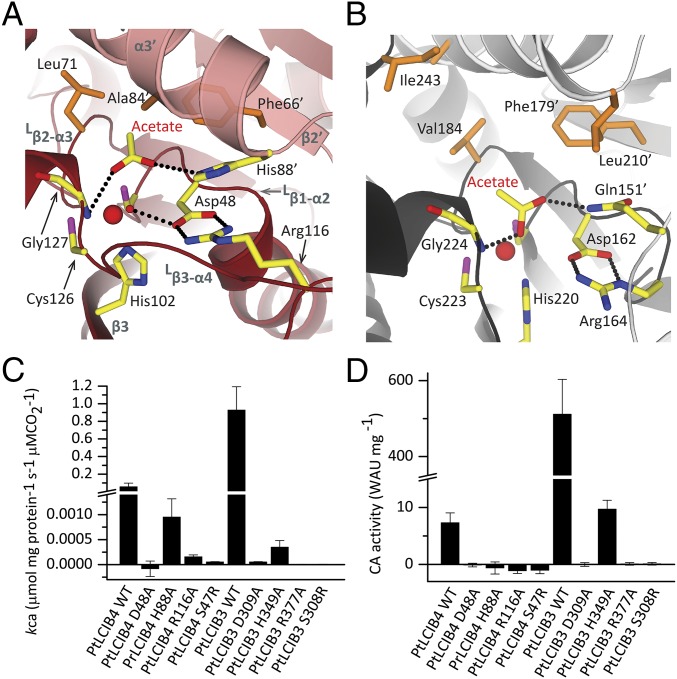
Fig. 4.
Detailed view of the active site in PtLCIB4 and PSCA, and activity assay. (A and B) The detailed view of the active site in PtLCIB4 (A) and PSCA (B). The two subunits of PSCA from pea P. sativum are colored dark gray and light gray. The side chains of the polar residues at the active site of the two structures are shown as yellow sticks. The hydrophobic residues involved in acetate binding at the active site are shown as orange sticks. (C and D) MIMS (C) and Wilbur–Anderson (D) CA activity assays of PtLCIB4, PtLCIB3, and their mutants are shown. Error bars indicate the mean and SD of at least three independent repeats.

