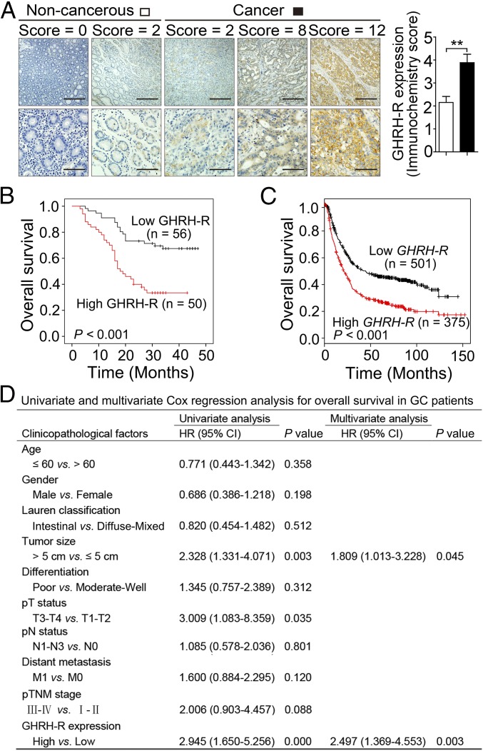
Fig. 1.
GHRH-R overexpression in GC patients is associated with poor survival. (A, Left) Immunohistochemical staining of GHRH-R (brown) on GC sections (n = 106) and paired noncancerous tissues. Nuclei were counterstained with hematoxylin (blue). (Scale bars: Top, 200 μm; Bottom, 50 μm.) (Right) The immunohistochemistry score of GHRH-R in GC (filled bar) and paired noncancerous (open bar) tissues was plotted. Error bars indicate SEM. **P < 0.01 by paired t test. (B) Kaplan–Meier curves compared the overall survival in GC patients with high and low protein levels of GHRH-R. (C) The relationship between overall survival and mRNA levels of GHRH-R in GC by Kaplan–Meier survival analysis. (D) Univariate and multivariate Cox regression analysis of clinicopathological factors in GC patients (n = 106).

