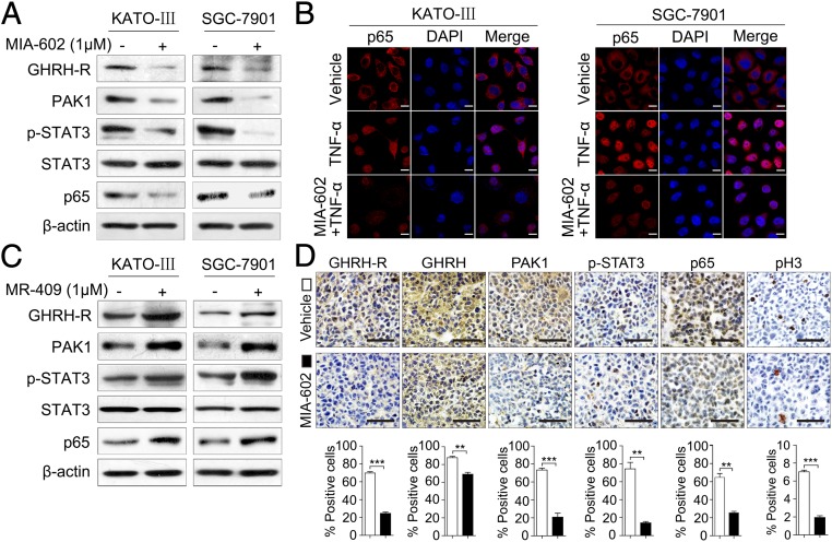
Fig. 4.
MIA-602 suppresses inflammatory signaling in GC. (A) KATO-III and SGC-7901 cells were treated with MIA-602 or vehicle and then were harvested, and equal amounts of lysates were immunoblotted for the labeled antigens. β-Actin was used as an internal control. (B) Subcellular localization of p65 (red) in the KATO-III and SGC-7901 cells, as analyzed by an immunofluorescence confocal assay. Nuclei were stained with DAPI (blue). (C) KATO-III and SGC-7901 cells were treated by MR-409 (1 μM) or vehicle and then were harvested. Equal amounts of lysates were immunoblotted for the labeled antigens. β-Actin was used as an internal control. (D, Upper) Representative images of immunohistochemistry of GHRH-R, GHRH, PAK1, p-STAT3, p65, and pH3 in tumors derived from SGC-7901 cells. (Lower) The percentages of treated (filled bars) and untreated (open bars) cells expressing the indicated proteins were plotted. Error bars indicate SEM. **P < 0.01, ***P < 0.001 by Student’s t test; n = 12 in each group. (Scale bars: B, 10 μm; D, 50 μm.)

