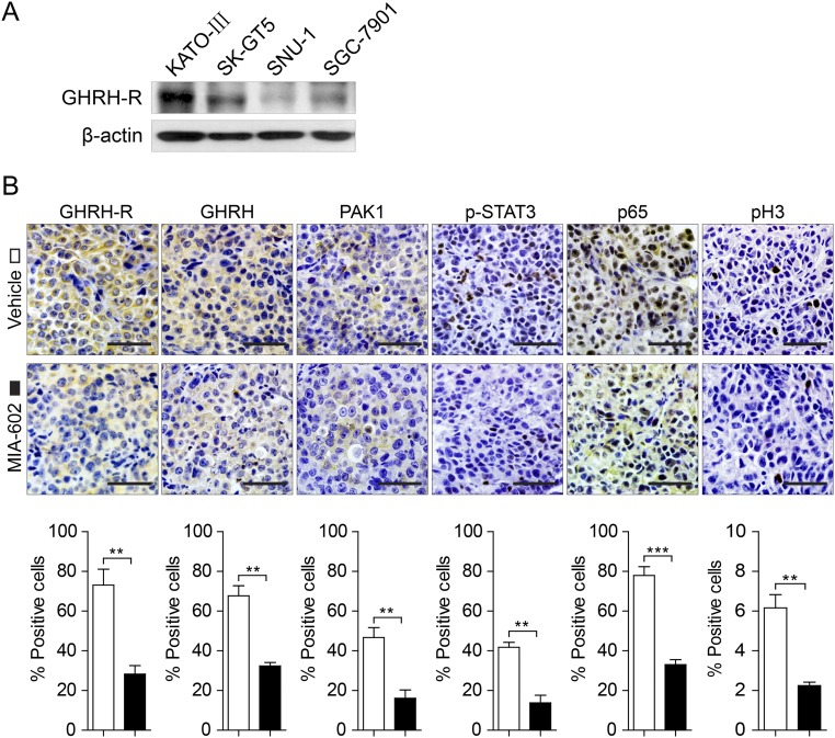
Fig. S4.
Expression of GHRH-R in GC cells and effects of MIA-602 on inflammatory signaling in vivo. (A) The status of GHRH-R in four GC cell lines was analyzed by Western blotting. β-Actin was used as an internal control. (B, Upper) Representative images of immunohistochemistry of GHRH-R, GHRH, PAK1, p-STAT3, p65, and pH3 in tumors derived from SK-GT5 cells treated with MIA-602. (Scale bars: 50 μm.) (Lower) The percentages of treated (filled bars) and untreated (open bars) cells expressing the indicated proteins were plotted. Error bars indicate SEM. **P < 0.01, ***P < 0.001 by Student’s t test; n = 12 in each group.

