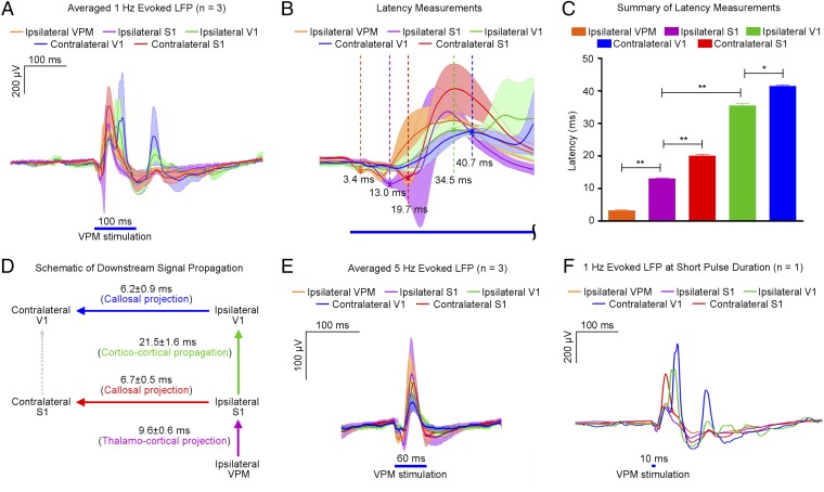
Fig. 5.
LFP recordings in sensory cortices and VPM reveal downstream signal propagation supported by low frequency (1 Hz) over long-range projections and their interactions with long temporal windows. (A) Averaged evoked LFPs during 1 Hz of VPM optogenetic stimulation (n = 3; 10% duty cycle; error bar indicates ± SD). (B) Summary of propagation delays from the onset of stimulation (magnified from A). Latency is measured as the time between the onset and the first evoked LFP peak location, indicated by a cross (x). (C) Summary of latency measured at the five different locations (n = 3; error bar indicates ± SD, one-way ANOVA with post hoc Bonferroni correction; **P < 0.01; *P < 0.05). (D) Schematic diagram of downstream signal propagation at 1 Hz. Optogenetic stimulation of VPM thalamo-cortical neurons elicited action potentials that excited ipsilateral S1 through thalamo-cortical projections. Subsequently, low-frequency activity propagated cortico-cortically from ipsilateral S1 to ipsilateral V1, and interhemispheric callosal projections between ipsilateral and contralateral S1 were recruited. Signal then was propagated from ipsilateral V1 to contralateral V1 via interhemispheric callosal projections. (E) Averaged evoked LFPs during 5 Hz of VPM optogenetic stimulation (n = 3; 30% duty cycle; error bar indicates ± SD). Comparision with 1 Hz reveals the absence of secondary peaks in ipsilateral and contralateral V1. This indicates that long-lasting and recurring interactions between and within cortico-cortical and thalamo-cortico-thalamic networks require long interstimulus durations. (F) Evoked potentials from a representative animal during 1 Hz of VPM optogenetic stimulation using a short stimulation pulse (1% duty cycle) shows no significant differences in overall latencies and LFP temporal characteristics.

