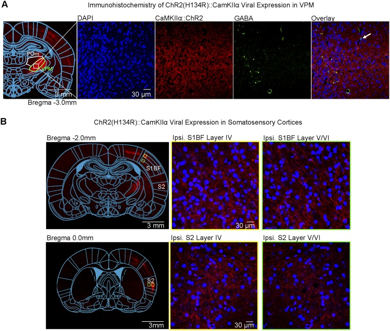
Fig. S1.
Histological characterization of ChR2::CaMKIIα viral expression in VPM thalamo-cortical excitatory neurons demonstrates no colocalization with VPM GABAergic neurons and their projection targets to S1BF and S2. (A) Confocal images of ChR2-mCherry expression in VPM; Lower magnification (Left) and higher magnification (Right). Overlay of images costained for the nuclear marker DAPI, mCherry, and inhibitory marker GABA revealed no colocalization between mCherry and VPM GABAergic neurons (indicated by white arrow). (B) Confocal images of ChR2-mCherry expression in S1BF (Top). Lower magnification (Left) and higher magnification (Right). VPM thalamo-cortical projections synapse in layer IV and V/VI of S1BF, indicated by no colocalization between mCherry and DAPI. Confocal images of ChR2-mCherry expression in S2 (Bottom) Lower magnification (Left) and higher magnification (Right). VPM thalamo-cortical projections synapse in layer IV and V/VI of S2, indicated by no colocalization between mCherry and DAPI.

