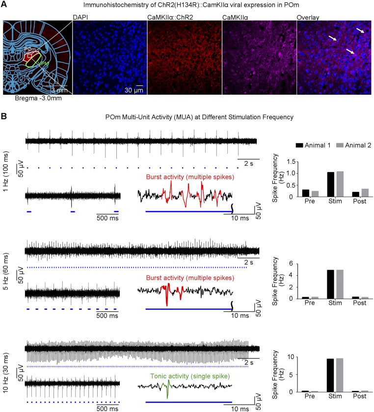
Fig. S5.
Histological characterization of ChR2::CaMKIIα viral expression in POm, and MUA recordings of POm thalamo-cortical excitatory neurons. (A) Confocal images of ChR2-mCherry expression in POm. Lower magnification (Left) and higher magnification (Right). Overlay of images costained for the nuclear marker DAPI, excitatory marker CaMKIIα, and mCherry revealed colocalization of mCherry and CaMKIIα in the cell body of POm thalamo-cortical neurons (indicated by white arrows). ChR2::CaMKIIα viral expression was shown to express only in POm without overlapping VPM. (B) MUA measured during POm optogenetic stimulation at three frequencies. Twenty-one-second trace of POm MUA from a representative animal during POm stimulation (1 Hz, 10% duty cycle; 5 Hz and 10 Hz, 30% duty cycle: Top Left to Bottom Left). Corresponding 2-s POm MUA trace magnified at the beginning of stimulation (Bottom Left) and illustration of optogenetically evoked thalamic burst (multiple spikes) or tonic activity (single spike; Bottom Right) for each stimulation frequency. Analysis of evoked spikes revealed an onset latency of 4.3 ± 2.3 ms (mean ± SD), consistent with POm thalamo-cortical neuron characteristics. Burst activity was elicited at 1 Hz and 5 Hz and transitioned to tonic activity at 10 Hz. Spike frequency measurements from two animals (Top Right to Bottom Right). They showed that spikes were successfully evoked by optogenetic pulses at all stimulation frequencies.

