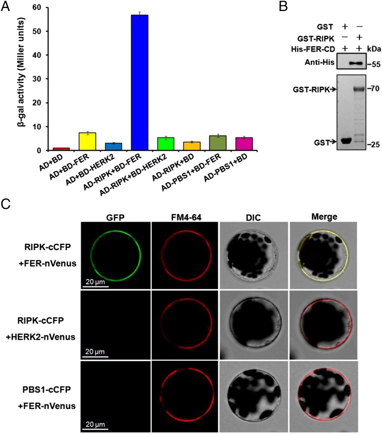
Fig. 1.
RIPK interacts with FER. (A) β-galactosidase assay in the Y2H system. AvrPphB susceptible protein 1 (PBS1) and HERKULES Receptor Kinase 2 (HERK2) were used as negative controls. RIPK and PBS1 were cloned into the pGADT7 vector (AD-RIPK, AD-PBS1) and FER-CD and HERK2-CD into the pGBKT7 vector (BD-FER, BD-HERK2). Experiments were repeated three times with similar results. (B) GST pull-down assay. Input GST (27 kDa, 2 μg) or GST-RIPK (70 kDa, 2 μg) protein was visualized by Coomassie Brilliant Blue staining (Lower). The eluted proteins were separated by a SDS/PAGE gel and probed with anti-His antibody (1:5,000) (Upper). Experiments were performed three times with similar results. (C) BiFC assay of FER–RIPK interaction in Arabidopsis protoplasts. Before imaging, the protoplasts were treated with FM4-64 (2 µM) for 5 min. GFP fluorescence was detected in protoplasts coexpressing RIPK-cCFP and FER-nVenus (Upper) but not in the controls (RIPK-cCFP + HERK2-nVenus or PBS1-cCFP + FER-nVenus; Middle and Lower). (Scale bar, 20 μm.)

