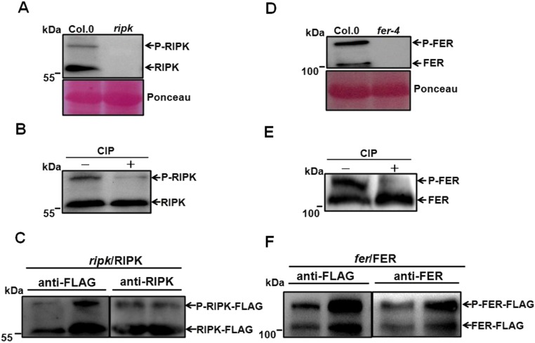
Fig. S5.
Specificity of FER and RIPK antibodies. (A) Anti-RIPK antibody recognized two bands in total protein extract from Col.0 but not ripk mutant. (B) RIPK has two forms: Without CIP treatment, the upper band is phosphorylated form (we named P-RIPK), and the lower band is nonphosphorylated form (we named RIPK), prominent after CIP treatment. (C) Specificity analysis of anti-RIPK antibody. Total protein was extracted from leaves of 7-d-old ripk/RIPK-FLAG complemented lines and probed with the anti-RIPK antibody (1:3,000) and anti-FLAG antibody (1:5,000), respectively. (D) Total protein was extracted from leaves of 45-d-old Col.0 and fer-4 mutant plants and was probed using the FER antibody (1:4,000) in a Western blot procedure. (E) FER was present in two forms: without CIP treatment, the upper band is phosphorylated form (we named P-FER), and the lower band is nonphosphorylated form (we named FER), prominent after CIP treatment. (F) Specificity analysis of anti-FER antibody using fer/FER complementation lines expressing FER-FLAG in the wild-type Col.0 background. Total protein was extracted from leaves of 35-d-old FER-FLAG plants and analyzed using the anti-FER antibody (1:4,000) and anti-FLAG antibody (1:5,000), respectively.

