Fig. 2.
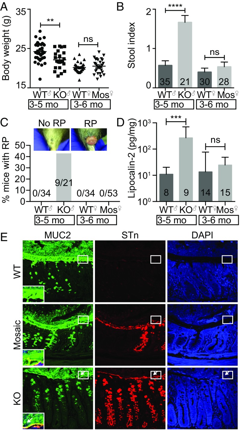
Spontaneous inflammation in IEC-Cosmc-KO but not mosaic mice. Body weight (n = 35 WT males, 21 KOs; 30 WT females, 28 mosaics) (A), stool index (average of softness and blood content) (B), and rectal prolapse for KO, mosaics, and gender matched WTs (C). (D) Fecal lipocalin-2 in KO, mosaics, and gender-matched WTs. (E) Distal colon from WT, mosaic, and KO were fixed with Carnoy’s reagent and stained with antibodies against MUC2 (green), STn (red), or with DAPI (blue) (3 mice per group, representative images shown); white boxes indicate the inner mucus layer, enlarged and merged in lower left corner; white arrows show where the inner mucus layer was lost. (Magnification: 40×.) **P ≤ 0.01; ***P ≤ 0.001; ****P ≤ 0.0001; ns, not signficant from two-tailed Student’s t test (A) or Mann–Whitney (B and D); mean ± SE (A and B) or median ± interquartile range (D); number of mice are indicated on the graph (B–D).

