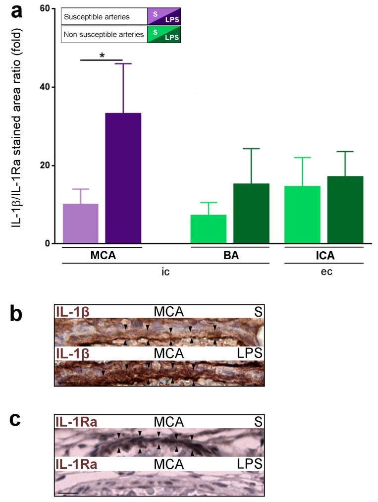
Figure 6.
IL-1β/IL-1Ra pro-inflammatory ratio in LPS-exposed (LPS group) vs. S-exposed (S group) P1 pups: (a) increased IL-1β/IL-1Ra pro-inflammatory ratio in LPS-exposed (LPS group) vs. S-exposed (S group) PAIS-susceptible artery; (b) IL-1β staining (black arrowhead) in a LPS-exposed vs. S-exposed PAIS-susceptible artery (MCA); and (c) IL-1Ra staining (black arrowhead) in a LPS-exposed vs. S-exposed PAIS-susceptible artery (MCA). Data are presented as mean ± standard error of the mean (SEM). * p < 0.05, Mann–Whitney test. n = 6–14 arteries from 6–7 animals per condition. Scale bar = 15 µm. Abbreviations: BA, basilar artery; ec, extra-cranial; ic, intra-cranial; ICA, internal carotid artery; IL-1β, interleukin-1β; LPS, lipopolysaccharide; MCA, middle cerebral artery; S, saline.

