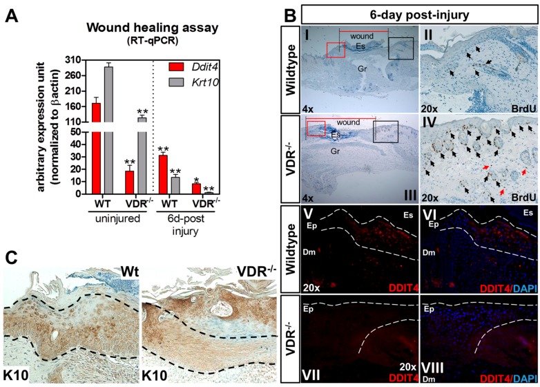
Figure 5.
Reduction of Ddit4 in the re-epithelized wounds of VDR−/− animals (column 1.5). (A) RT-qPCR analysis of the wound repair process from excised wounds. For uninjured samples, comparisons were made between genotypes. Six days following injury, sample comparisons were made between the respective uninjured genotypes. One-way ANOVA at an α = 0.05 (95% confidence interval) and Tukey’s multiple comparison post-tests were utilized. Significance is denoted with asterisks: * p < 0.05, ** p < 0.01 (n = 4 samples per genotype and time point); (B) Representative slides showing 5-bromo-2′-deoxyuridine (BrdU) immunohistochemical (I–IV) and Ddit4 immunofluorescence (V–VIII) staining of WT and VDR−/− skins six days following injury. Wound closure is depicted by the horizontal red bars. Black boxed areas in the 4× slides represent the BrdU-labeled magnified regions (II and IV). Red-boxed areas represent the Ddit4-labeled magnified regions (V–VII). In the BrdU slides, black arrows depict proliferating epidermal and follicular keratinocytes. Red arrows depict proliferating dermal fibroblasts. In the Ddit4 panels, the dotted white line marks the re-epithelialized area. Nuclei are marked in blue with 4′,6-diamidino-2-phenylindole (DAPI), while Ddit4 is labeled in red. ES: eschar; GR: granulation tissue; EP: epidermis; DM dermis; (C) Krt10 immunostaining within the neo-epidermis (demarcated by dashed lines) six days following injury.

