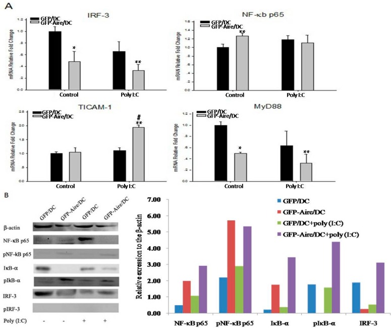
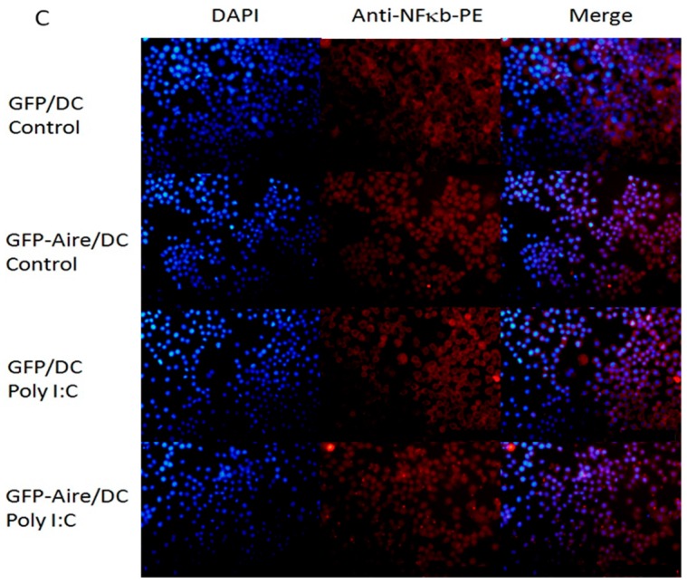
Figure 4.
Aire modifies key TLR3 pathway molecules in DC2.4 cells. (A) The IRF-3, NF-κB, TICAM-1, and MyD88 transcript expression levels were detected in GFP-Aire/DC and GFP/DC by RT-qPCR. All qPCR data are shown as the expression calculated relative to that of GAPDH and are depicted as fold changes relative to the expression in GFP/DC cells, which was normalized to 1; (B) NF-κB, IκB-α, and IRF-3 protein expression levels were measured by Western blotting. The histograms are the relative expression of each molecules compared to the β-actin; and (C) Immunofluorescent detection of NF-κB (red) translocation to the nuclei (blue; combined signal, purple) in DC2.4 cells. Original magnification, 200×. A representative experiment of three independent experiments is shown. Data are shown as the means ± SD from three to six independent experiments. * p < 0.05 and ** p < 0.01 compared with the GFP/DC control; # p < 0.05 compared with the GFP-Aire/DC control.


