Figure 1.
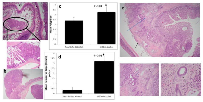
Light:dark (LD) shift accelerates intestinal tumorigenesis in alcohol treated mice: (a) Tubular adenoma in the ileum (4×). The circle is a 10× view of the same lesion, showing clear delineation between adenoma (right) and adjacent normal mucosa (left); (b) Colonic tubular adenoma (4×); (c) Polyp size (* p = 0.01) and (d) number of large polyps (* p = 0.01) were increased significantly by light:dark shifting; (e) Only mice in the experimental group developed invasive colon cancer; picture shows invasive adenocarcinoma emerging from a tubular adenoma. Two glands immediately above muscularis mucosa (MM) (MM is indicated by the blue arrow) are infiltrating the mucosa, and a few glands in top center have infiltrated the submucosa (top circle). Area of invasion to MM and submucosa is also observed in the center-bottom (blue circle). Part of the polyp surface was eroded—an inconsequential finding (black arrow). Right-bottom panel is a larger view (10×) of the same polyp, showing glands infiltrating into the MM (blue arrow) and beyond (black arrow). The left-bottom panel is the high power view (40×) of two glands that have infiltrated into the submucosa. At this power, desmoplasia is apparent as a subtle rim around the glands (black arrow); desmoplasia is an unequivocal feature of invasive carcinomas. Note: 4×, 10×, and 40× fields are 25.0 microns, 10.0 microns, and 2.5 microns in diameter, respectively.

