Figure 1.
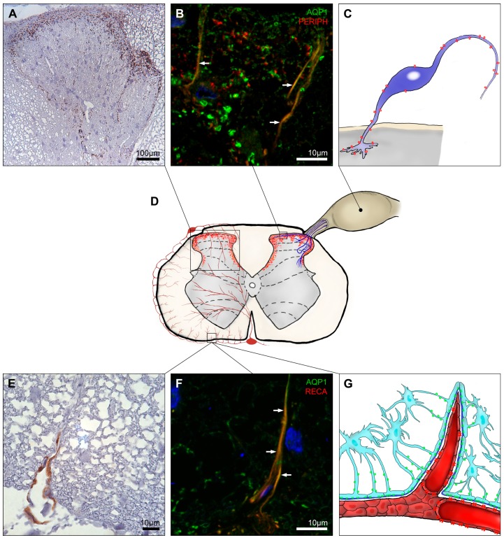
AQP1 expression in the spinal cord. AQP1 was strongly expressed at laminae I and II of the dorsal horn with decreasing signal intensity at the medial edges of dorsal horns up to lamina V (A,D); A fraction of the AQP1 signal belonged to unmyelinated neuronal cells (B) protruding from dorsal root ganglion to the superficial laminae of dorsal horns which axons build up peripheral sensory fibers (C); Illustration of AQP1 expression in the spinal cord cross-section (D); AQP1 labeling in the white matter was rather infrequent and found in proximity to the glia limitans (E) most likely belonging to small arterioles furcating from the arterial vasocorona (F,G). Panels A, B, E and F are modified from Oklinski et al. [21]. The blue signal in panels B and F represents 4′,6-diamidino-2-phenylindole (DAPI) staining of the nuclei; AQP1, aquaporin 1; PERIPH, peripherin and RECA, rat endothelial cell antigen-1. Colocalization indicated by arrows. AQP1 is indicated in red on panels C, D and G. AQP4 is drawn in green on panel G. White arrows in B and F.

