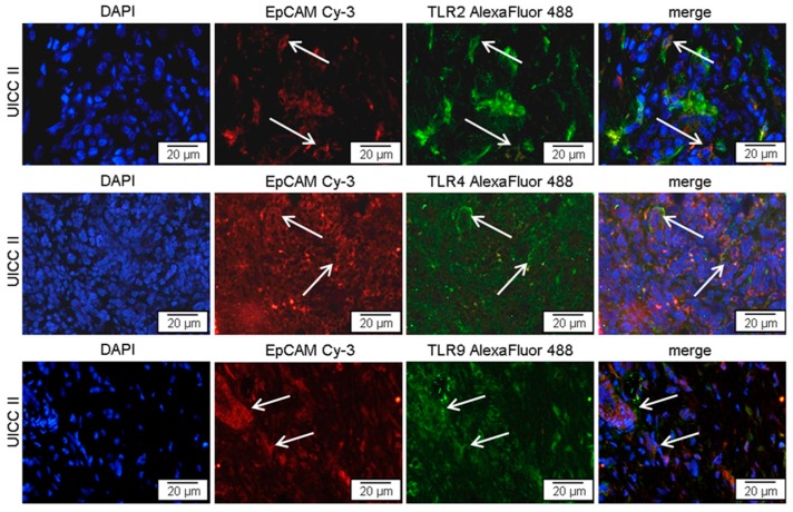
Figure 2.
TLR2, -4, or -9 expressing tumor cells in pancreatic cancer tissue. Representative examples of immunofluorescence double staining, showing TLR (green) and EpCAM (red) co-staining (arrows) in tumor cells of patients with pancreatic cancer UICC II. AlexaFluor 488, green; Cy3 (indocarbocyanin), red; DAPI (49,6-diamidino-2-phenylindoldihydrochlorid), blue—nuclear counterstaining.

