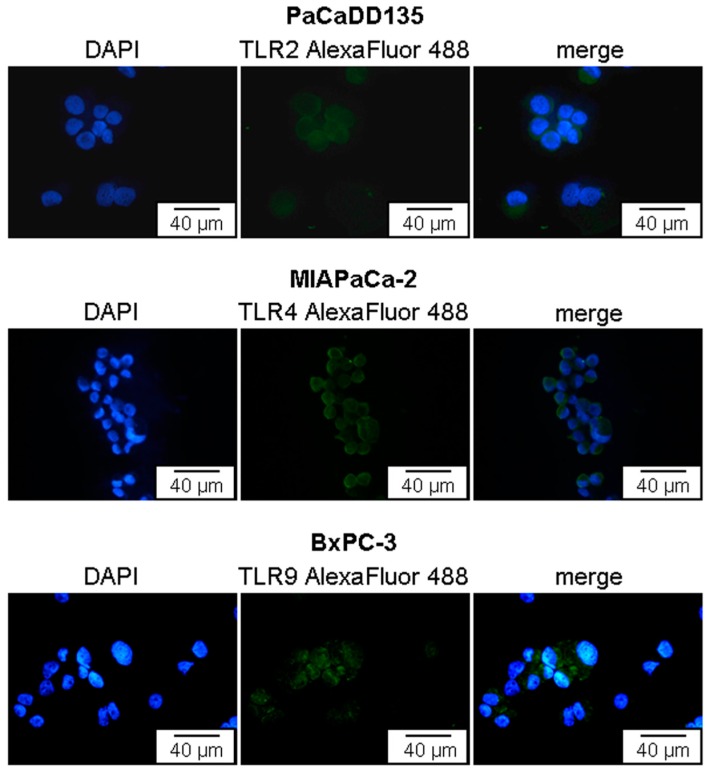
Figure 4.
Immunofluorescent staining of TLR2, -4, and -9 in PaCaDD135, MIAPaCa-2, and BxPC-3 pancreatic cancer cells. Representative examples showing TLR2, -4, and -9 expression (green). Cell surface localization of TLR2 and -4 as well as intracellular localization of TLR9 can be identified. AlexaFluor 488, green; DAPI (49,6-diamidino-2-phenylindoldihydrochlorid), blue—nuclear counterstaining.

