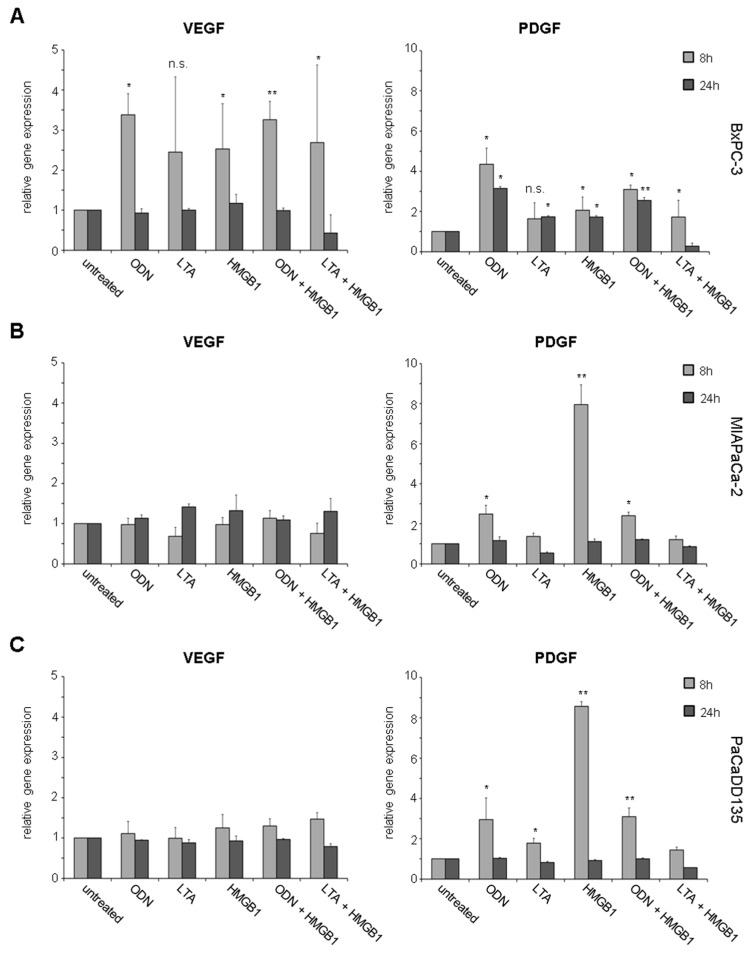
Figure 6.
Gene expression analysis of VEGF and PDGF in pancreatic cancer cell lines after treatment with TLR ligands: BxPC-3 (A); MIAPaCa-2 (B); and PaCaDD135 (C) were incubated with ODN, LTA, HMGB1, ODN + HMGB1, and LTA + HMGB1 and analyzed by RT-qPCR 8 and 24 h after stimulation. Increased VEGF gene expression was observed only in BxPC-3 cells, whereas PDGF was significantly expressed in all three cancer cell lines. Untreated cells were standardized to baseline. The relative gene expression is expressed as 2−ΔΔCq. * p < 0.05, ** p < 0.005, n.s. not significant.

