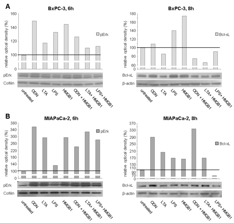
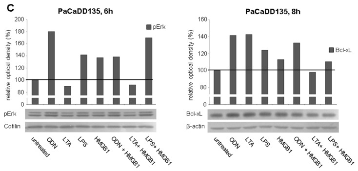
Figure 8.
Western blot analysis of pErk and Bcl-xL in different human pancreatic cancer cells after treatment with TLR ligands: BxPC-3 (A); MIAPaCa-2 (B); and PaCaDD135 cells (C) were incubated with ODN, LTA, LPS, HMGB1, ODN + HMGB1, LTA + HMGB1, and LPS + HMGB1 and analyzed 6 h (pErk) and 8 h (Bcl-xL) after stimulation. Cofilin (pErk) and β-actin (Bcl-xL) probes were used as controls for protein loading. Relative optical density (ROD) was determined using ImageJ software. Values for proteins of interest were calculated in relation to values of loading controls. Untreated cells were standardized to baseline.


