
Fig. 2.
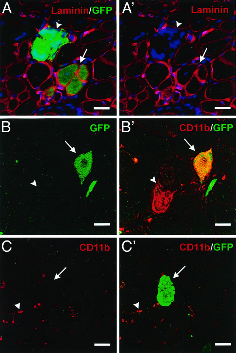
Injected BMDC contribute to intact myofibers or give rise to CD11b/Mac1+ cells that invade degenerating fibers. (A and A′) Representative confocal microscopic analyses of transverse sections of skeletal muscle 4 weeks after i.m. injection of cells fractionated from the BM of GFP transgenic mice detect GFP (green in A only), laminin (A and A′, red) and nuclei (A and A′, blue). Both GFP+ myofibers surrounded by basal lamina (arrow) and GFP+ clusters of cells not surrounded by basal laminal membranes (arrow heads) were observed in TA cross sections (note number of nuclei in GFP+ cluster in A′). (B and B′) GFP+ clusters (arrows) (green) were positive for CD11b (B′, red) and therefore identified as donor-derived macrophages that have invaded degenerating myofibers. Endogenous macrophages (arrowheads) are not GFP+.(C and C′) True GFP+ myofibers (arrows) (C′, green) were found to lack CD11b (red), whereas infiltrating endogenous macrophages CD11b+ (arrowheads) were observed. (Scale bars, 50 μm.)