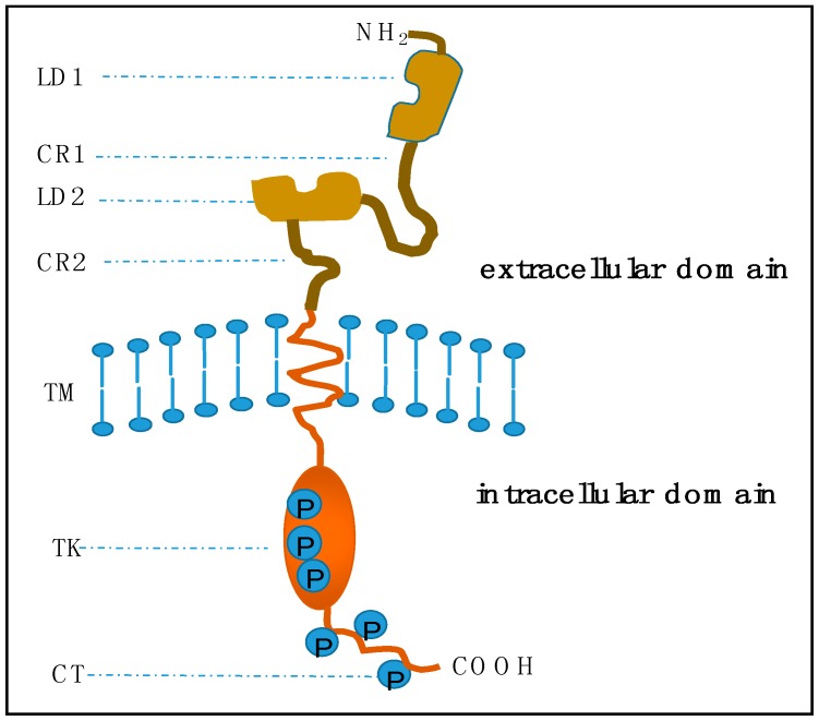
Figure 1.
Basic structure of epidermal growth factor receptor (EGFR) transmembrane proteins. In the extracellular domain, LD1 and LD2 are two repeated ligand binding domains. CR1 and CR2 are two repeated cysteine rich regions. TM indicates the short transmembrane spanning sequences. In the intracellular domain, TK is a catalytic tyrosine kinase, and CT is the carboxyl-terminal tail. Circled Ps are the phosphorylation sites within the TK and CT regions. This figure is revised based on the review of the oncogene human epidermal growth factor 2 (HER2) contributed by Moasser [17].

