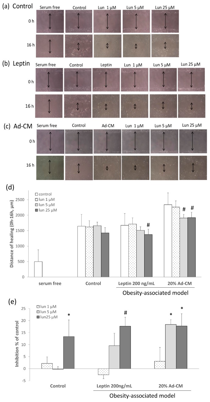
Figure 2.
Effect of lunasin on breast cancer 4T1 cell metastasis. Migration patterns were observed in the scraped area of 4T1 cells treated with or without lunasin for 16 h and incubated under three conditions: (a) Fresh medium-5% fetal bovine serum/Dulbecco’s modified Eagle’s medium (FBS/DMEM), the mark of double arrows was present the distance of healing in the pictures; (b) Leptin at 200 ng/mL in 5% FBS/DMEM; (c) 20% Ad-CM in 5% FBS/DMEM; (d) The distance of migrated cells was quantified by manual counting under the microscope scale; (e) Migrated cells were quantified by manual counting under the microscope and presented as a percentage of inhibition relative to that of control (control as 0%). Data are shown as mean ± SEM. Statistical analysis was tested by one-way ANOVA and then Fisher’s LSD test; significant differences are presented as * p < 0.05, or # p < 0.01 versus control group.

