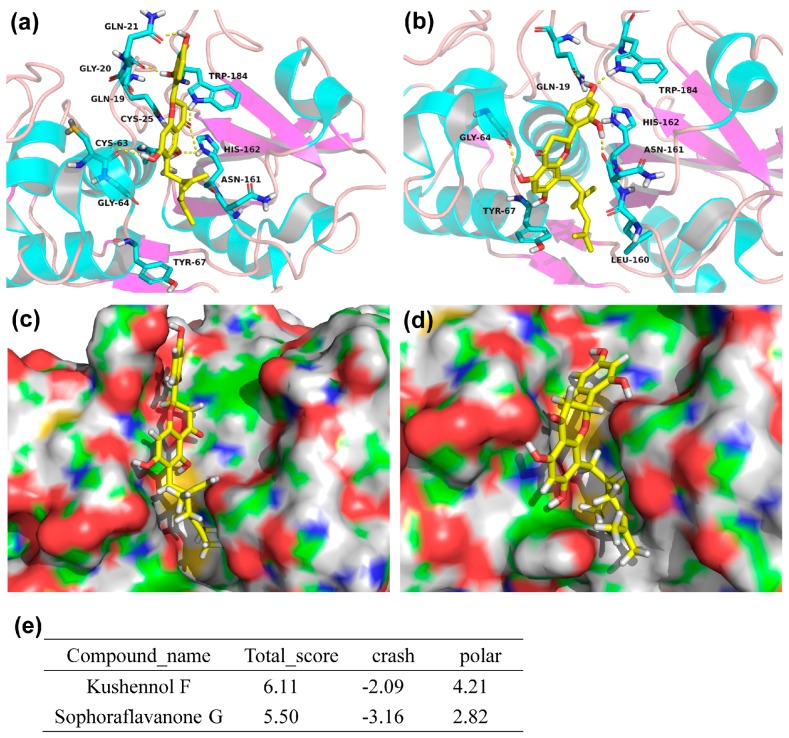
Figure 6.
Predicted binding mode of KF and SG to the active site of Ctsk (PDB entry: 3KWZ). The structures of compounds were represented in stick model with color (yellow: carbon atom, red: oxgen atom and white: hydrogen atom). The binding mode of KF (a) and SG (b) in the active site of Ctsk. The protein was shown in the cartoon representation, the key residues in the active site were also represented as stick model with color (blue, red, and gray), the yellow dashed line denoted protein-ligand H-bonding interactions. The docking pose of KF (c) and SG (d) located in active site of Ctsk. The protein was illustrated by a solid surface. (e) The Docking Scores as acquired by Surflex-dock.

