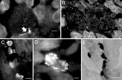
Fig. 1.
Immunostaining of kidney and heart with anti-renin antibody. (A) The polyclonal anti-renin Ab (1:500) exhibits specific binding to rat kidney at the vascular pole of the glomerulus (arrow). (Scale bar = 10 μm.) (B) No staining was seen in sections exposed to the polyclonal anti-renin Ab (1:500) preadsorbed with an excess of human renin. (Scale bar, 10 μm.) (C and D) Staining of sections of rat ventricle prepared with the polyclonal anti-renin Ab (1:500) and viewed with a ×40 (C) or ×100 (D) objective. (Scale bars, 10 μmin C and 4 μmin D.) (E) Staining of mast cells in rat ventricle with toluidine blue. (Scale bar, 20 μm.)

