Fig. 2.
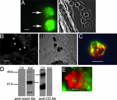
Presence of immunoreactive renin in cardiac mast cells. (A and A1) Costained section of rat ventricle with the polyclonal anti-renin Ab (A) and toluidine blue (A1). (Scale bar, 5 μm.) (B and B1) A section of rat ventricle prepared with the polyclonal anti-renin Ab (1:500) preadsorbed with an excess of human renin (B) and stained with toluidine blue (B1). The toluidine-blue-stained mast cells (B1) did not immunoreact with the preadsorbed anti-renin Ab (see asterisks in B). (C) A cardiac mast cell colabeled with the monoclonal anti-renin Ab (red) (1:100) and an antihistamine Ab (green) (1:500). (Scale bar, 10 μm.) Areas stained with both Abs appear yellow. (D) Western blot of rat kidney homogenate (Kid) (20 μg per lane) and pure CD (500 μg per lane) probed with the polyclonal anti-renin Ab (1:12,500) and an anti-CD Ab (1:100). (E) A cardiac mast cell colabeled with anti-CD Ab (green) (1:500) and the polyclonal anti-renin Ab (red) (1:500). (Scale bar, 5 μm.)

