Fig. 4.
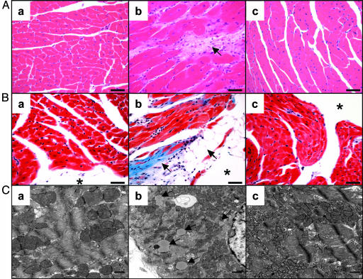
Comparison of the myocardial histology of wild-type (a), ACS-transgenic treated with AdCMV-β-gal (b), and ACS-transgenic treated with AdCMV-leptin (c) mice. (A) Hematoxylin/eosin stain showing myofiber disorganization, cardiomyocyte enlargement, and interstitial fibrosis in the AdCMV-β-gal-treated ACS-transgenic group but a normal appearance in the hyperleptinemic group. (Bar, 40 μm.) (B) Trichrome stain of the hearts showing collagen deposition in the subendocardium and interstitium; cells resembling adipocytes can be seen near the lumen of the heart. The hyperleptinemic ACS-transgenic group is entirely normal. (Bar, 40 μm.) (C) Electron microscopic appearance of myocardial cells of the three groups. Lipid vacuoles in cardiomyocytes of the AdCMV-β-gal-treated ACS-transgenic mice are marked by arrows. None are noted in the other two groups. (* marks the lumen of the heart). (Bar, 500 nm.)

