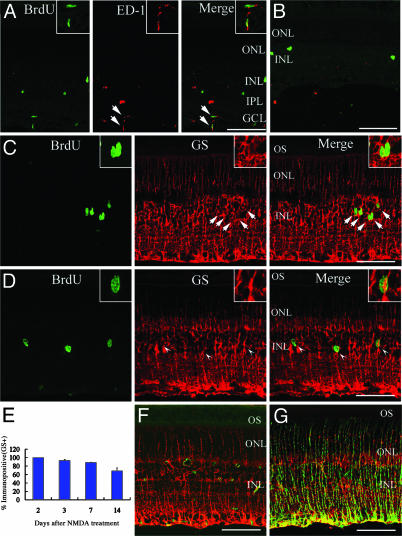
Fig. 2.
Müller glia proliferate, become active, and convert to other cell types. (A) At DAN2, some BrdUrd-labeled cells (green) in the GCL and IPL expressed ED-1 (red). (B) At DAN14, most BrdUrd-labeled cells (green) did not express ED-1 (red). (C) At DAN2, all BrdUrd-labeled cells (green) in the INL expressed GS (red). Note that GS labeling is detected from GCL to ONL, but not in OS (outer segments of photoreceptors). (D) At DAN3, some BrdUrd-labeled cells (green) did not express GS (red). Arrowhead, BrdUrd-positive/GS-negative cell. (Inset) Magnified view. (E) Percentage of BrdUrd-labeled cells in the INL that expressed GS at different times after NMDA treatment. Data are mean ± SD obtained from at least four animals. (F) Sections of retina treated with PBS and labeled with nestin (green) and GS (red). (G) Sections of retina obtained at DAN2 and labeled with nestin (green) and GS (red). (Insets in A and C) Magnified view of double labeling cells (arrows). (Scale bar, 100 μm.)

