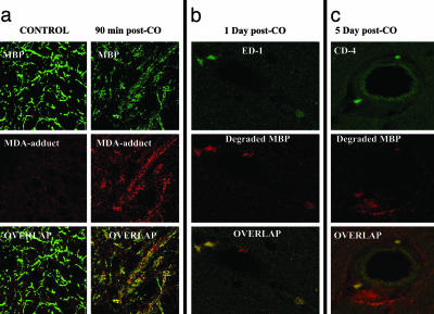
Fig. 1.
Immunohistochemical evaluations. (a) Control brain section showing low-level staining by antibody to MDA-protein adducts but no colocalization with native MBP, and extensive staining for MDA-adducts with colocalization to MBP (shown in yellow in “overlap” images) in brains of rats killed 90 min after CO poisoning. (b) Colocalization among the microglia/macrophage marker, ED-1, and “degraded MBP” at 1 day after CO poisoning. (c) Infiltration of CD-4+ lymphocytes observed in close proximity to an accumulation of “degraded MBP” at 5 days after CO poisoning.

