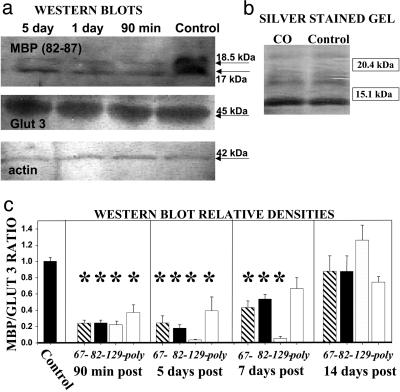
Fig. 3.
Western blots of MBP after CO poisoning. (a) Representative Western blot probed with a monoclonal antibody that recognizes the MBP epitope between amino acids 82 and 87, GLUT 3, and actin-using brain homogenates from rats killed at different times after CO exposure. (b) Silver stain of SDS/PAGE gel from a control rat and one killed 90 min after CO poisoning. (c) Quantification of MBP staining expressed as the ratio of band densities for MBP versus GLUT 3 with four different antibodies to MBP (monoclonal antibodies to amino acid segments 67–74, 82–87, and 129–138, and a polyclonal anti-MBP). Blots were generated for rats killed at different times after poisoning. Data are expressed relative to a control ratio of 1.0. Homogenates from five different rats were used for all blots probed. Values are mean ± SE. *, P < 0.05.

