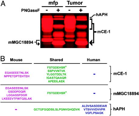
Fig. 2.
General MS-based strategy to distinguish stromal (mouse) and carcinoma (human) enzyme activities in xenograft tumors. (A) Expanded view of representative FP-labeled tumor and mfp SH activities showcasing the increased resolution that is achieved for glycosylated enzyme activities (e.g., mCE-1) after treatment with peptide: N-glycosidase F (PNGaseF). Identities of enzyme activities are shown on either side of the gel: hAPH, human acyl-peptide hydrolase; and mMGC18894, uncharacterized mCE. (B) Tryptic peptide maps for representative SH activities shown in A. Blue, peptides unique to human orthologue; magenta, peptides unique to mouse orthologue; green, peptide shared between mouse and human orthologues. The asterisk highlights a peptide that is shared between CE-1 and MGC18894 enzymes.

